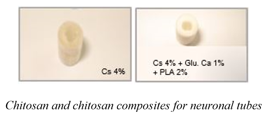
- Neuronal tubes,
- chitosan,
- calcium glutamate,
- polylactid acid,
- freezer-dried
Copyright (c) 2020 SChQ

This work is licensed under a Creative Commons Attribution-NonCommercial-ShareAlike 4.0 International License.
Abstract
Nerve regeneration is a biologically complex phenomenon. Once the nervous system is damaged, its recovery is difficult and causes a malfunction in other parts of the body that can occur because mature neurons do not suffer cell division. To increase the prospects of axonal regeneration and functional recovery, research has focused on the construction and design of nerve conduction channels or guide tubes. That is why a high molecular weight chitosan was used as a starting material, which was first characterized by the following analyses: moisture, ash, FTIR, TGA, total nitrogen determination, viscosity for the determination of the average molecular weight and degree of deacetylation by potentiometric titration. Once the starting chitosan is characterized, tube-shaped biomaterials are prepared based on a 4% w/v chitosan solution, using CH3COOH solvent at 1% v/v. Calcium glutamate is added to 0.5% and 1% w/v, neutralizing with NaOH 0.25N, and then taken to the molds to be frozen and subsequently freeze-dried. Finally, the materials are coated with polylactic acid, in order to achieve a strengthening in the structure of the material. These materials are also prepared in a bio clean area. The different materials are chemically and morphologically characterized by FTIR, TGA, Atomic Absorption Spectroscopy, Mechanical Properties, and Morphology and solubility test.
References
- Afifi AK. Neuroanatomía functional. Texto y Atlas. México (DF): McGraw–Hill Interamericana, (1999).
- Catala M , Teillet M -A , De Robertis E M , Le Douarin N M . A spinal cord fate map in the avian embryo: While regressing, Hensen's node lays down the notochord and floor plate thus joining the spinal cord lateral walls. Development, 122:2599–2610, (1996).
- Moury J D , Schoenwolf G C . Cooperative model of epithelial shaping and bending during avian neurulation: Autonomous movements of the neural plate, autonomous movements of the epidermis, and interactions in the neural plate/epidermis transition zone. Dev. Dyn, 204: 323–337, (1995).
- Smith J L , Schoenwolf G C . Neurulation: Coming to closure. Trends Neurosci, 11:510–517, (1997).
- Vassilis EK, Rajiv RR. Cell death and diseases of the nervous system. Totowa, Nueva Jersey: Humana Press, (1986).
- Garbay B, Heape, AM, Sargeil, F, Cassage C. Myelin synthesis in the peripheral nervous system. Prog Neurobiol, 61:267–304, (2000).
- Smith KS, Kapoor R, Hall SM, Davies M. Electrically Active Axons Degenerate When Exposed to Nitric Oxide. Ann Neurol, 49:470–476, (2001).
- Araki T, Milbrandt J. Ninjurin, A novel adhesion molecule, is induced by nerve injury and promotes axonal growth. Neuron, 17:353–361, (1996).
- Perry V, Luner E, Brown M. Evidence that three rate of Wallerian degeneration is controlled by a single autosomal dominant gene. Eur J Neurosci, 2:402–408, (1990).
- Evanthia Nikolopoulou, Gabriel L. Galea, Ana Rolo, Nicholas D. E. Greene, Andrew J. Copp
- Neural tube closure: cellular, molecular and biomechanical mechanisms. Development, 144: 552-566; (2017). doi: 10.1242/dev.145904
- Chaudry V, Glass J, Friffin J. Wallerian degeneration in peripheral nerve disease. Neurol Clin, 10:613–627, (1992).
- Dumitru, D. Reaction of the peripheral nervous system to injury. In electrodiagnostic medicine. Filadelfia: Ed Hanley–Belfus Inc, (1994).
- Weiss P. The technology of nerve regeneration. Sutureless tubulation and related methods of nerve repair. Neurosurgery, 1:400– 450, (1944).
- Ayala H. Estudio Experimental en la cirugía de los nervios periféricos. Rev Ortop Traum, 2:3–14, (1997).
- Sulaiman ARO, Gordon T. Effects of Short– and Long–Term Schwann Cell. Denervation on Peripheral Nerve Regeneration, Myelination, and Size. Glia, 32: 234–246, (2000).
- Barakat W. The role of thyroid hormones and their receptors in peripheral nerve regeneration. J Neurobiol, 40:541, (1999).
- Dahlin LB., Lundborg G. Use of tubes in peripheral nerve repair. Neurosurgery Clinics of North America, 1:341-352, (2001).
- G. Sundback C, Hadlock T, Cheney M, Vacanti J. Biomaterials; 24: 819 – 830, (2003).
- Benavides-Cuellar, MA. Quitina-Quitosana. Los polímeros del futuro. División de comunicaciones, Servicio Nacional de Aprendizaje SENA, Centro de Desarrollo Tecnológico y Asistencia Técnica a la Industria. Santiago de Cali, (2002).
- Aguado A, David S, Richardson P, Bray G. Axonal elongation in peripheral and central nervous system transplants. Adv Cell Neurobiol, 3:215, (1982).
