SYNTHESIS OF AG0 NANOPARTICLES FROM EXTRACT AND ETHANOLIC AND AQUEOUS FRACTIONS OF LEAVES AND PETALS OF HIBISCUS ROSA-SINENSIS L.
- Green synthesis,
- Hibiscus rosa-sinensis L.,
- FE-SEM,
- UHPLC,
- Nanoparticles
- EDS,
- DLS,
- Ultrasound-assisted extraction ...More
Copyright (c) 2022 SChQ

This work is licensed under a Creative Commons Attribution-NonCommercial-ShareAlike 4.0 International License.
Abstract
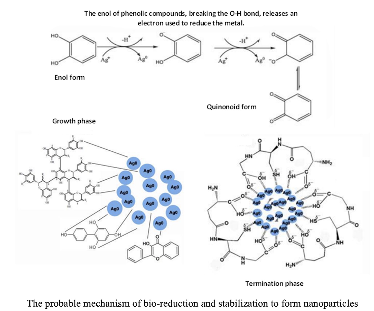
The synthesis of nanoparticles from plant extracts has become an interesting line of research in recent years. The purpose of the study was the synthesis of silver nanoparticles (NSP) from silver nitrate, aqueous and ethanol 80% extracts obtained by ultrasound-assisted extraction, as well as fractions of chloroform, ethyl acetate and water, of flower petals and leafs of Hibiscus rosa-sinensis L. The ethanolic extract of petals of H. rosa-sinensis L. was analyzed by UHPLC-ESI-Q-Orbitrap-MS/MS identifying pelargonidin, petunidin, kaempferol, luteolin and orientin. Characterization of NSP by UV-Visible spectrophotometry gave a lmax at 400,631 nm and 389,411 nm for NSP obtained with ethanolic extract of flower petals and leafs, lmax at 402,270 nm and 391,057 nm for NSP of aqueous extracts of flower petals and leafs respectively. FTIR confirmed the reduction of Ag+ ions to Ag0 ions in NSP. Dynamic light dispersion (DLS) showed an effective diameter of the NSP from extracts, less than 80 nm and for NSP from fractions was less at 53 nm and near-zero polydispersity indices. Electron field emission scanning microscopy (FE-SEM) showed a particle size between 17 - 32 nm for NSP from extracts, and 15 - 26 nm for NPS from fractions, which showed a spherical structure. Energy dispersion X-ray spectroscopy (EDS) confirmed the elemental composition of NSP showing mostly silver (60.44%), oxygen (32.48%) and potassium (4.97%). This study revealed that the compounds from the extracts of Hibiscus rosa-sinensis L. are good reducing and stabilizing agents for the synthesis of silver nanoparticles, being pH 9 optimal for synthesis.
References
- S. Singh, K. C. Barick, and D. Bahadur, “Surface engineered magnetic nanoparticles for removal of toxic metal ions and bacterial pathogens,” J. Hazard. Mater., vol. 192, no. 3, pp. 1539–1547, 2011, doi: 10.1016/j.jhazmat.2011.06.074.
- W. R. Li, X. B. Xie, Q. S. Shi, H. Y. Zeng, Y. S. Ou-Yang, and Y. Ben Chen, “Antibacterial activity and mechanism of silver nanoparticles on Escherichia coli,” Appl. Microbiol. Biotechnol., vol. 85, no. 4, pp. 1115–1122, 2010, doi: 10.1007/s00253-009-2159-5.
- H. M. M. Ibrahim, “Green synthesis and characterization of silver nanoparticles using banana peel extract and their antimicrobial activity against representative microorganisms,” J. Radiat. Res. Appl. Sci., vol. 8, no. 3, pp. 1–11, 2015, doi: 10.1016/j.jrras.2015.01.007.
- M. del R. Sarmiento, E. Ortiz, and J. Alvarez, “Emergencias ambientales asociadas a sustancias químicas en México,” Gac. Ecológica, Secr. Medio Ambient. y Recur. Nat., no. 66, pp. 54–63, 2003.
- P. Dong et al., “The green synthesis of Ag-loaded photocatalyst via DBD cold plasma assisted deposition of Ag nanoparticles on N-doped TiO2 nanotubes,” J. Photochem. Photobiol. A Chem., vol. 382, no. March, p. 111971, 2019, doi: 10.1016/j.jphotochem.2019.111971.
- S. Ahmed, M. Ahmad, B. L. Swami, and S. Ikram, “REVIEW A review on plants extract mediated synthesis of silver nanoparticles for antimicrobial applications : A green expertise,” J. Adv. Res., vol. 7, no. 1, pp. 17–28, 2016, doi: 10.1016/j.jare.2015.02.007.
- Y. W. Mak, L. O. Chuah, R. Ahmad, and R. Bhat, “Antioxidant and antibacterial activities of hibiscus ( Hibiscus rosa-sinensis L .) and Cassia ( Senna bicapsularis L .) flower extracts,” J. King Saud Univ. - Sci., vol. 25, no. 4, pp. 275–282, 2013, doi: 10.1016/j.jksus.2012.12.003.
- D. Philip, “Green synthesis of gold and silver nanoparticles using Hibiscus rosa sinensis,” Phys. E Low-dimensional Syst. Nanostructures, vol. 42, no. 5, pp. 1417–1424, 2010, doi: 10.1016/j.physe.2009.11.081.
- S. M. Seyyednejad, H. Koochak, E. Darabpour, and H. Motamedi, “A survey on Hibiscus rosa-sinensis, Alcea rosea L. and Malva neglecta Wallr as antibacterial agents,” Asian Pac. J. Trop. Med., pp. 351–355, 2010, doi: 10.1016/S1995-7645(10)60085-5.
- R. Patel, A. Patel, S. Desai, and A. Nagee, “Study of Secondary Metabolites and Antioxidant Properties of Leaves , Stem and Root among Hibiscus Rosa-Sinensis cultivars,” ASIAN J. EXP. BIOL. SCI., vol. 3, no. 4, pp. 719–725, 2015.
- Z. A. Khan et al., “Antioxidant and antibacterial activities of Hibiscus Rosa-sinensis Linn flower extracts,” Pak. J. Pharm. Sci., vol. 27, no. 3, pp. 469–474, 2014.
- R. Sharmila Devi and R. Gayathri, “Green Synthesis of Zinc Oxide Nanoparticles by using Hibiscus rosa-sinensis,” Int. J. Curr. Eng. Technol., vol. 4, no. 4, pp. 2444–2446, 2014.
- D. Nayak, S. Ashe, P. R. Rauta, and B. Nayak, “Biosynthesis, characterisation and antimicrobial activity of silver nanoparticles using Hibiscus rosa-sinensis petals extracts,” IET Nanobiotechnology, vol. 9, no. 5, pp. 288–293, 2015, doi: 10.1049/iet-nbt.2014.0047.
- E. Aguilar and P. Bonilla, “Actividad antioxidante e inmunológica de flavonoides aislados de hojas de Smallanthus sonchifolius (Yacón),” Cienc. Invest., vol. 12, no. 1, pp. 15–23, 2009, [Online]. Available: http://scholar.google.com/scholar?hl=en&btnG=Search&q=intitle:Actividad+antioxidante+e+inmunol�gica+de+flavonoides+aislados+de+hojas+de+SMALLANTHUS+SONCHIFOLIUS+(YAC�N)#0.
- G. Rangel and P. Adriana, “Cuantificación de flavonoides ( Catequinas ) en cáscara de naranja variedad criolla ( Citrus sinensis ) producida en Norte de Santander,” LIMENTECH Cienc. Y TECCNOLOGÍA Aliment., vol. 8, no. 2, pp. 34–43, 2010.
- O. Torres, A. Angulo, M. Montaño, and P. Galeano, “ESTUDIO QUIMICO Y OBTENCION DE PRINCIPIOS ACTIVOS DE LA ESPECIE Rollinia,” no. 33, pp. 55–58, 2007.
- O. R. Lock, INVESTIGACIÓN FITOQUÍMICA: Métodos en el estudio de productos naturales. 2016.
- R. M. Ibrahim, A. M. El-Halawany, D. O. Saleh, E. M. B. El Naggar, A. E. R. O. EL-Shabrawy, and S. S. El-Hawary, “HPLC-DAD-MS/MS profiling of phenolics from securigera securidaca flowers and its anti-hyperglycemic and anti-hyperlipidemic activities,” Brazilian J. Pharmacogn., vol. 25, no. 2, pp. 134–141, 2015, doi: 10.1016/j.bjp.2015.02.008.
- N. A. Begum, S. Mondal, S. Basu, R. A. Laskar, and D. Mandal, “Biogenic synthesis of Au and Ag nanoparticles using aqueous solutions of Black Tea leaf extracts,” Colloids Surfaces B Biointerfaces, vol. 71, no. 1, pp. 113–118, 2009, doi: 10.1016/j.colsurfb.2009.01.012.
- F. N. Domenech Gordillo, “Síntesis y caracterización de nanopartículas de plata usando extracto de hojas de Ambrosia arborescens (marco) como reductor químico,” 2017.
- R. Zanella, “Metodologías para la síntesis de nanopartículas : controlando forma y tamaño,” Mundo Nano, vol. 5, no. 1, pp. 69–81, 2012, doi: http://dx.doi.org/10.22201/ceiich.24485691e.2012.1.45167.
- S. Ahmed, S. Ullah, M. Ahmad, and B. L. Swami, “Green synthesis of silver nanoparticles using Azadirachta indica aqueous leaf extract,” J. Radiat. Res. Appl. Sci., vol. 9, no. 1, pp. 1–7, 2015, doi: 10.1016/j.jrras.2015.06.006.
- A. T. Madrid Sani, “SÍNTESIS Y CARACTERIZACIÓN DE NANOPARTÍCULAS DE PLATA A PARTIR DE VARIOS EXTRACTOS PIGMENTADOS DE DOS PLANTAS PARA SU APLICACIÓN EN CELDAS SOLARES HÍBRIDAS,” 2017.
- I. Hussain, N. B. Singh, A. Singh, H. Singh, and S. C. Singh, “Green synthesis of nanoparticles and its potential application,” Biotechnol. Lett., vol. 38, no. 4, pp. 545–560, 2016, doi: 10.1007/s10529-015-2026-7.
- C. T. Du and F. J. Francis, “ANTHOCYANINS OF ROSELLE (Hibiscus sabdariffa, L.),” J. Food Sci., vol. 38, no. 5, pp. 810–812, 1973, doi: 10.1111/j.1365-2621.1973.tb02081.x.
- A. Gärtner, G. Gellerstedt, and T. Tamminen, “Determination of phenolic hydroxyl groups in residual lignin using a modified UV-method,” Nord. Pulp Pap. Res. J., vol. 14, no. 2, pp. 163–170, 1999, doi: 10.3183/npprj-1999-14-02-p163-170.
- M. Barragán Condor, J. M. Aro Aro, A. E. Muñoz Cáceres, and J. Rodríguez Mendoza, “Determinacion de antocianinas y capacidad antioxidante en extractos de (Muehlembeckia volcanica),” Rev. Investig. Altoandinas - J. High Andean Res., vol. 22, no. 2, pp. 161–169, 2020, doi: 10.18271/ria.2020.604.
- C. J. Vargas Fajardo, “DETERMINACIÓN Y CUANTIFICACIÓN DE COMPUESTOS FENOLICOS EN FLORES DE TARAXACUM OFFICINALE, MEDIANTE HPLC-DAD-MS Y ENSAYOS COLORIMETRICOS UV-VIS.,” 2020.
- H. M. Al-Yousef, W. H. B. Hassan, S. Abdelaziz, M. Amina, R. Adel, and M. A. El-Sayed, “UPLC-ESI-MS/MS Profile and Antioxidant, Cytotoxic, Antidiabetic, and Antiobesity Activities of the Aqueous Extracts of Three Different Hibiscus Species,” J. Chem., vol. 2020, 2020, doi: 10.1155/2020/6749176.
- J. A. M. Fuentes, L. López-Salas, I. Borrás-Linares, M. Navarro-Alarcón, A. Segura-Carretero, and J. Lozano-Sánchez, “Development of an Innovative Pressurized Liquid Extraction Procedure by Response Surface Methodology to Recover Bioactive Compounds from Carao Tree Seeds,” Foods, vol. 10, no. 2, p. 398, 2021, doi: 10.3390/foods10020398.
- T. G. García Morales, M. Á. Vásquez Guevara, and S. Lagunas Rivera, “Extracción de antocianinas en los cálices de Hibiscus sabdariffa L. como posible fotosensibilizador.,” p. 6, 2019.
- M. Goutam et al., “Authentication and Photochemical Screening of Hibiscus Rosa Sinensis .,” IJRAR, vol. 5, no. 4, pp. 712–718, 2018.
- J. P. Pérez-Orozco, L. M. Sánchez-Herrera, E. Barrios-Salgado, and M. T. Sumaya-Martínez, “Kinetics of solid-liquid extraction of anthocyanins obtained from Hibiscus rosa-sinensis,” Rev. Mex. Ing. Química, vol. 12, no. 3, pp. 505–511, 2020, [Online]. Available: http://www.redalyc.org/articulo.oa?id=62029966013.
- A. Purushothaman, P. Meenatchi, S. S, R. Sundaram, and N. Saravanan, “Quantification of Total Phenolic Content, HPLC Analysis of Flavonoids and Assessment of Antioxidant and Anti-haemolytic Activities of Hibiscus rosa-sinensis L. Flowers in vitro,” Int. J. Pharma Res. Heal. Sci., vol. 4, no. 5, pp. 134–50, 2016, doi: 10.21276/ijprhs.2016.05.02.
- H. Tran Trung, H. Truong Thi Huynh, L. Nguyen Thi Thuy, H. Nguyen Van Minh, M. N. Thi Nguyen, and M. N. Luong Thi, “Growth-inhibiting, bactericidal, antibiofilm, and urease inhibitory activities of hibiscus rosa sinensis L. flower constituents toward antibiotic sensitive- And resistant-strains of helicobacter pylori,” ACS Omega, vol. 5, no. 32, pp. 20080–20089, 2020, doi: 10.1021/acsomega.0c01640.
- H. Z. Lina, M. M. Samy, A. E. B. Samir, A. M. Fatma, M. T. Kawther, and A. S. Abdelaaty, “Hypoglycemic and antioxidant effects of Hibiscus rosa-sinensis L. leaves extract on liver and kidney damage in streptozotocin induced diabetic rats,” African J. Pharm. Pharmacol., vol. 11, no. 13, pp. 161–169, 2017, doi: 10.5897/ajpp2017.4764.
- C. Y. Kuo, E. S. Kao, K. C. Chan, H. J. Lee, T. F. Huang, and C. J. Wang, “Hibiscus sabdariffa L. extracts reduce serum uric acid levels in oxonate-induced rats,” J. Funct. Foods, vol. 4, no. 1, pp. 375–381, 2012, doi: 10.1016/j.jff.2012.01.007.
- L. P. Guan and B. Y. Liu, “Antidepressant-like effects and mechanisms of flavonoids and related analogues,” Eur. J. Med. Chem., vol. 121, pp. 47–57, 2016, doi: 10.1016/j.ejmech.2016.05.026.
- N. Vasudeva and S. K. Sharma, “Biologically active compounds from the genus Hibiscus,” Pharm. Biol., vol. 46, no. 3, pp. 145–153, 2008, doi: 10.1080/13880200701575320.
- H. Y. Chai, S. M. Lam, and J. C. Sin, “Green synthesis of magnetic Fe-doped ZnO nanoparticles via Hibiscus rosa-sinensis leaf extracts for boosted photocatalytic, antibacterial and antifungal activities,” Mater. Lett., vol. 242, pp. 103–106, 2019, doi: 10.1016/j.matlet.2019.01.116.
- S. Surya, G. Dinesh Kumar, and R. Rajakumar, “Green Synthesis of Silver Nanoparticles from Flower Extract of Hibiscus rosa-sinensis and Its Antibacterial Activity,” Int. J. Innov. Res. Sci. Eng. Technol. (An ISO, vol. 5, no. 4, pp. 5242–5247, 2016, doi: 10.15680/IJIRSET.2016.0504129.
- A. Santos-Espinoza, F. Gutiérrez-Miceli, V. Ruíz-Valdiviezo, and J. Montes-Molina, “El papel de los compuestos polifenólicos en la síntesis verde de nanopartículas metálicas,” BioTecnología, vol. 24, no. 2, p. 46, 2020.
- K. Vijayaraghavan and S. P. K. Nalini, “Biotemplates in the green synthesis of silver nanoparticles,” Biotechnol. J., vol. 5, no. 10, pp. 1098–1110, 2010, doi: 10.1002/biot.201000167.
- D. Garg, A. Shaikh, A. Muley, and T. Marar, “In-vitro antioxidant activity and phytochemical analysis in extracts of Hibiscus rosa-sinensis stem and leaves,” Free Radicals Antioxidants, vol. 2, no. 3, pp. 41–46, 2012, doi: 10.5530/ax.2012.3.6.
- A. Shehzad et al., “Synthesis, characterization and antibacterial activity of silver nanoparticles using Rhazya stricta,” PeerJ, vol. 2018, no. 12, pp. 1–15, 2018, doi: 10.7717/peerj.6086.
- K. Vijayaraghavan and T. Ashokkumar, “Plant-mediated biosynthesis of metallic nanoparticles: A review of literature, factors affecting synthesis, characterization techniques and applications,” J. Environ. Chem. Eng., vol. 5, no. 5, pp. 4866–4883, 2017, doi: 10.1016/j.jece.2017.09.026.
- K. Muthu and S. Priya, “Green synthesis, characterization and catalytic activity of silver nanoparticles using Cassia auriculata flower extract separated fraction,” Spectrochim. Acta - Part A Mol. Biomol. Spectrosc., vol. 179, pp. 66–72, 2017, doi: 10.1016/j.saa.2017.02.024.
- N. Chandra Lekha and M. Selvipriya, “Green Synthesis of Silver Nanoparticles from aqueous leaves extract of Hibiscus rosa- sinensis and its antioxidant activity,” Kamaraj J. Acad. Res., vol. 1, no. 3, pp. 21–28, 2018.
- M. M. H. Khalil, E. H. Ismail, K. Z. El-baghdady, and D. Mohamed, “Green synthesis of silver nanoparticles using olive leaf extract and its antibacterial activity,” Arab. J. Chem., vol. 7, no. 6, pp. 1131–1139, 2014, doi: 10.1016/j.arabjc.2013.04.007.
- R. Herrera Basurto, B. Simonet Suas, and M. Valcárcel Cases, “Retos de las mediciones en la nanoescala,” in Simposio de metrología, 2012, pp. 1–6.
- A. E. Al-Snafi, “Arabian medicinal plants with analgesic and antipyretic effects-plant based review (Part 1),” IOSR J. Pharm., vol. 8, no. 6, pp. 81–102, 2018.
- D. Alvear, S. Galeas, and A. Debut, “Síntesis y Caracterización de Nanopartículas de Magnetita,” Rev. Politécnica, vol. 39, no. 2, pp. 61–66, 2017, doi: 10.33333/rp.v39i2.545.
- S. Ahmed, Annu, S. A. Chaudhry, and S. Ikram, “A review on biogenic synthesis of ZnO nanoparticles using plant extracts and microbes: A prospect towards green chemistry,” J. Photochem. Photobiol. B Biol., vol. 166, pp. 272–284, 2017, doi: 10.1016/j.jphotobiol.2016.12.011.
- L. Katata-Seru, T. Moremedi, O. S. Aremu, and I. Bahadur, “Green synthesis of iron nanoparticles using Moringa oleifera extracts and their applications: Removal of nitrate from water and antibacterial activity against Escherichia coli,” J. Mol. Liq., vol. 256, pp. 296–304, 2018, doi: 10.1016/j.molliq.2017.11.093.
